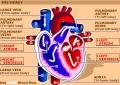
The above picture shows the different regions of the heart. Heart is a muscular organ in our body, that is made of four chambers in humans as we as in all mammals. In fishes and amphibians though, it has been observed to be of three chambers. The muscles are of special type, called the cardiac muscles. They have inter-fibrillar branches that simply acts as functional synctitium, having gaph junctions. This helps the heart muscles to contract circumferentially as a whole, as the pace maker stimulus passes down the line of action.
Heart is autorhythmic in nature. It means that it beats on its own, and that too in a rythmical pattern. This rythmical pattern is enabled by some special kinds of tissues called the pace maker tissues. There are four different types found within the cardiac muscular mass: 
1) Sino Atrial Node (SAN)
2) Atrio Ventricular Node (AVN)
3) Bundle of His
4) Purkinjee Fibers.
The figure in the adjacent plate shows the orientation of these tissues. The SAN is located at the entry point of teh Superior Vena Cava of the right atrium. Three fibers attach SAN to the AVN. These fibers aids in transfer of some electrical potential to the AVN, but some also passes through the atrial walls, as a result of which Atrail Systole (systole= contraction) occurs. From the AVN, the impulse next passes to the tip of the ventricles, via the Bundle of His, through the Intraventricular septum. The AVN helps in delaying the transfer of the impulse. This is significant because, the delay causes a proper emptying of the blood from the atrial chambers into the respective Ventricles. The next plate would show the movement of blood in the whole cycle. The Bundle of His enables the impulse to move to the tip, by the previously mentioned path. This also has some significance. The impulse is liberated onto the Ventricular Walls and hence Ventricular Systole occurs, and the systolic movement ot the contractile movement must be from the tip to the auriculo-ventriculo valves. This enables the blood to close the two septal valves(Mitral Valve and Bicuspid Valve) and pushes the blood from the Ventricles to respective large arteries, namely, Aorta by Left Ventricle and Pulmonary Artery by Right Ventricles.
As mentioned earlier, the pace maker tissue is able to cause suc potentials because it can alter the ionic concentration within the tissue. There is a major Calcium and Sodium influx(entry) by means of certain Calcium and Sodium channels. While there is a non equivalent Pottassium efflux. These alters the ionic charges and hence produces a propagatory action potential. It can be recorded in ECG (Electro Cardio Graph) recordings.
p.s: ECG and Blood movement pictures to be posted later on. The inconvinience is regretted.
------------Somnath Paul.
email: somnath4you@gmail.com